
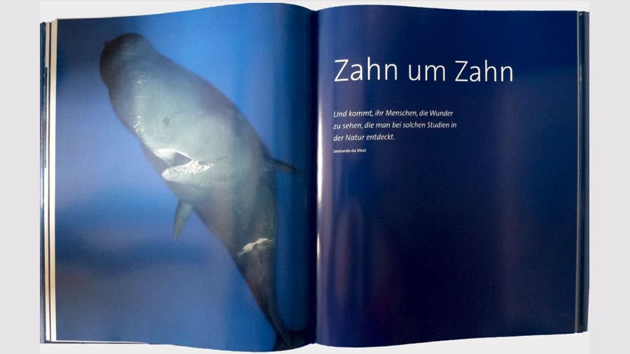
Whales – Up Close
Excerpt from our chapter: moments
The eye

They are blue, brown, gray or green. Our eyes can shine, cry, shine or even hypnotize.
For us humans, the eyes are the most important sensory organ, as they are responsible for around 70 percent of our daily perception
Despite all the perfection, the human eye cannot achieve the charisma of a whale's eye, because anyone who has ever looked deeply into the eyes of a whale or dolphin never forgets these moments.
We feel awe, euphoria and admiration almost simultaneously. Also because we know that we are among the few people who have ever looked into the unfathomable eyes of these underwater giants.
But what is so special about them that they captivate us so much?
It can't be due to their size, because the eyes of whales and dolphins are rather small in relation to their body size. They are not one of the most important sensory organs, because the eyesight of all whales is not extremely well developed and is only roughly equivalent to that of deep-sea fish.
For some it is enough to just distinguish between light and dark, others are completely blind and all of them have severely limited color vision.
But how does “seeing” come about?
Seeing
In all eyes, the incident light rays are transmitted through the cornea, through the pupil and then via the lens to the retina. Receptors in this retina convert the light into electrical nerve impulses and send them via the optic nerve to the vision centers in the brain. Actual vision works in whales just like it does for us humans.
So do whales “see” the same things we do?
If we look deeply into the whales' eyes, we first discover many similarities with the human eye in the structure and structure of the eye.

The human eye
Our almost spherical eye, which is why it is also called an eyeball, has a diameter of approx. 2.5 cm and consists of three layers of tissue, as well as the lens and the vitreous body, which is filled with a gelatinous mass. The eyeball, surrounded by a lubricating fatty body, lies well protected in the eye sockets formed by the skull bones. The three layers of tissue form the wall of the eyeball. The first outer layer with the cornea and the sclera, the second with the choroid and iris and on the back part the third layer with the retina.
The outer dermis is whitish in color and is therefore also called “white eye skin”. If we look in the mirror, this white sclera can be clearly seen as the “white” in our eyes. It extends from the posterior entry point of the optic nerve, almost completely encloses the eyeball, merges into the transparent and avascular cornea at the front and has the task of keeping the eye in shape and protecting it. Between the dermis and the retina, on the second middle layer of tissue, lies the choroid, which, with its many blood vessels, supplies the adjacent layers with nutrients and oxygen. When we look in the mirror we can sometimes see the small red choroidal vessels shining through the “white” of our eyes.
A striking feature of the front section of the choroid is the iris, which is responsible for the color of our eyes. This is influenced by the pigment melanin. A high concentration of melanin results in brown eyes, while a lower pigment content makes the iris appear green, blue or gray, depending on the dosage.
In the middle of the iris is a round hole called the pupil. With the help of muscles, the iris can enlarge or shrink the pupil, thereby controlling how much light passes through the pupil onto the lens and the retina behind it.
When relaxed, our lens has the shape of an ellipse, is more flat and thin and in this shape regulates the sharpness of distance. Muscles that are connected to the lens and the eyeball enable us to change the shape of the lens through an interaction of tension and relaxation and thus create different degrees of curvature. When these muscles contract and the lens takes on a more rounded shape, we can see objects well at close range.
After being “focused”, the light rays bundled by the lens are passed on through the vitreous body to the inner tissue layer of the eyeball, to the retina, where an inverted, reduced image is initially created.
The retina then continues to work like an image editing program on the computer, because before the image is forwarded, the light receptors and the retinal nerve cells are responsible for displaying the movements in the image more clearly, improving the image contrast and making the colors brighter.
The retinal ganglion cells are also located in the retina, nerve cells whose projections act as “optic nerves” and carry the electrically encoded signals into the brain.
The light world ends at the retina.
Light therefore plays a very important role in our “seeing” so that we can orientate ourselves in our environment.
The whale eye

But despite their poor vision, the eyes of marine mammals are masterpieces of evolution. They have adapted excellently to the marine living conditions and we can discover amazing things in the eyes of the whales, because we can find cornea, sclera, lens, retina and much more, even tear fluid in their eyes.
Their protection is also adequately ensured, because the white outer skin of the eye, the dermis, is particularly thick in whales, almost rock hard, which is why it is also called the sclera, which is derived from the Greek “scleros = hard”, and the eye in front It is intended to protect against deformations, while the oily fluid from the tear glands protects the cornea.
Human eyes are almost spherical, although whale eyes are more like a hemisphere because they are noticeably flattened in the front area.

The light

The Lens

In the 1980s, scientists hypothesized that this shifting of the lens might be responsible for being able to see clearly in and out of water. However, through more recent research, they found that the cornea, with its unusual shape, probably helps the whales' eyes adapt well to seeing in water and in the air. This theory is also confirmed by the ophthalmologist and scientist Bert van der Pol from Groningen in the Netherlands, who specializes in the examination of marine mammal eyes, because he modeled a computer program and, with the help of the data from Kröger and Kirschfeld, combined it with his observations.
The cornea
He discovered that the center of the whale's cornea curves more inwards than outwards and is not uniformly thick. It is thinner in the middle and thicker towards the outside, but it is very flat, which certainly offers advantages under water, but in the air it means that the whales should be extremely short-sighted with up to minus 15 diopters.
Underwater, the marine mammals are more or less normal-sighted due to the round lens and this shape of the cornea, and in the air, this inwardly curved cornea acts like a strongly negatively refractive lens, which largely neutralizes myopia. The whale eye has, so to speak, negative contact lenses built into the cornea.
Almost all whales have thus solved the problem of myopia. However, this type of corneal change has not yet been demonstrated in studies of sperm whale eyes.

The sharpness
But not only the sperm whale, but also we humans do not have this ability of a built-in negative contact lens, because we have all had the experience that with our eyes open underwater everything appears blurred and blurry to us.
The reason for this lies in the different optical densities of water and air. In order to create a sharp image for us humans, the light is refracted on the surface of the cornea, which then passes it on to the lens. This in turn directs it to the retina, to the yellow spot, the point of sharp vision. The distance between the object, the lens of the eye and the retina must be in a certain relationship to one another.
By curving the lens, we can change the focal length and thus focus the distance between the object and the lens. However, due to the fixed lens size of the eye, the distance between the eye lens and the retina can never be changed.
Due to the higher density of water, light is refracted and scattered when air enters water. It hits the cornea more differently than in the air. Our lens now has to adapt much more to focus on the distance between the object and the lens. However, it only succeeds to a limited extent and as a result the image that it transmits to the retina is no longer transported at the correct distance. The resulting image ends up behind the retina and we see it blurry.
The whales have solved this problem with the so-called negative built-in contact lens. If we want to see clearly under water, we would need glasses with 40 diopters or we could simply put on diving goggles. This creates the normal density of air in front of the cornea and our usual refraction of light. We see clearly again.
The color
However, marine mammals are not only very nearsighted, they are even color blind compared to us humans and other mammals.
Around 127 million cells in the retina are responsible for our color and light vision. 120 million of these cells are so-called rods that are very sensitive to light and can only distinguish between light and dark. The other 7 million are called cones and are divided into three types. They respond to light in the blue, green and red wavelength range, which is why we also speak of trichomatic color vision. We are able to distinguish almost ten million shades from mixtures of these three basic colors.
Most mammals only have two types of cones, namely the blue and green cones. Some only have blue and red cones. This bichromatic color vision is, so to speak, the basic equipment of mammals. But two large groups of marine mammals, whales and seals, fall completely outside of this pattern.
Leo Peichl from the Max Plank Institute for Brain Research, Günther Behrmann from the Alfred Wegener Institute in Bremerhaven and Ronald Kröger from Sweden discovered an astonishing deficit in joint examinations of 14 different species of toothed whales, seals and sea lions. Whales and seals even lack blue cones. They only have one type of cone, namely the green cone. They are therefore cone monochromats and practically color blind.
The retina
For the sperm whales, the loss of the blue cones should be irrelevant anyway, because they spend most of their time diving and in the depths to which they penetrate, colors and light no longer play a major role.

However, in order to use the remaining light more efficiently even at shallower depths, the whales have developed a reflective mirror layer, the tapetum lucidum, which lies behind the retina. Undoubtedly, we are all familiar with the bright green eyes of a cat or dog when they are illuminated in the evening by the rays of a car lamp or the flash of a camera. This light-amplifying layer serves to capture the incident light and reflect it back, whereby the light passes through the retina again.
This back and forth improves light sensitivity because the sensory cells on the retina have a second chance to react to the incoming light quantum and thus utilize it better and more. Many nocturnal land mammals have this tapetum lucidum; the only difference in marine mammals is that theirs is a little grayer instead of green.
The tapetum covers at least the upper two-thirds of the fundus of the eye in all whales, and in some even the entire area. Such complete coverage of the fundus of the eye is unique among mammals in whales.
But the whales amazed the researchers even more, because two yellow spots were discovered on the whales' retina and thus two areas of sharpest vision, the macula lutea. The human eye only has one macula and this tiny point on our retina, with its 147,000 visual cells per mm2, is responsible for essential vision such as reading and recognizing details. The rest of the retina only primarily perceives outlines, speeds and contrasts of light and dark.

The sperm whale eye

The vitreous body was filled with an oily mass that is much tougher and denser than ours. The intraocular pressure in this animal could no longer be measured, because in dead animals it reaches zero within a few minutes, while in dead people it can take hours until it has scorched.
At the back of the eyeball, van der Pol was also able to expose the optic nerve, a huge bundle of nerve fibers that exits the retina. It reached an impressive length of almost one meter. In contrast, the human optic nerve only reaches 4.5 cm. Surprisingly, the 2.5 mm diameter of this optic nerve was only slightly larger than that of a human with its 1.5 mm. The optic nerve, as well as a large part of the back of the eye, was embedded in a supporting tissue that formed a dense, branched network of blood vessels and blood cavities, the so-called miracle network.

Über uns
- Kontakt
- Impressum
- Datenschutz
Nehmen Sie Kontakt auf.
Post-Adresse
Pottwale e.V.
Rotterdamer Straße 35
40474 Düsseldorf
+49 173-976 78 23
Bücher
- Pottwale - Im dunklen Blau des Meeres
Tauchen Sie ein in die faszinierende Welt der Pottwale
Pottwale sind auch in unserer computergesteuerten High-Tech-Zeit noch immer geheimnisvolle Wesen, von deren toten Körpern wir fast alles, von ihrem Leben aber so gut wie gar nichts wissen. Die sanften Riesen, deren Männchen bis zu 20 Meter groß werden können, leben im offenen Ozean und tauchen in unerforschte Tiefen. Sie kämpfen mit den Giganten der Tiefsee, deren Zeuge Suppenteller-große Merkmale an ihrer Haut sind. Keine andere Walart ist so bekannt wie der Pottwal, wurde so erbarmungslos gejagt und in so großer Zahl getötet.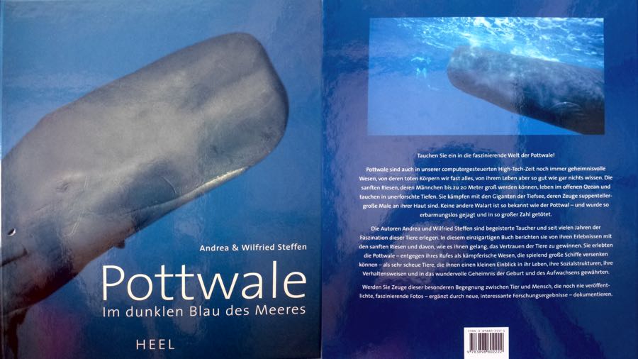 Die Autoren Andrea und Wilfried Steffen sind begeisterte Taucher und seit vielen Jahren der Faszination dieser Tiere erlegen. In diesem einzigartigen Buch berichten sie von Ihren Erlebnissen mit den sanften Riesen und davon, wie es ihnen gelang, das Vertrauen der Tiere zu gewinnen. Sie erlebten die Pottwale – entgegen ihres Rufes als kämpferische Wesen, die spielend große Schiffe versenken können- als scheue Tiere, die ihnen einen kleinen Einblick in ihr Leben, ihre Sozialstrukturen, ihre Verhaltensweisen und in das wundervolle Geheimnis der Geburt und des Aufwachsen gewährten.
 Werden Sie Zeuge dieser besonderen Begegnung zwischen Tier und Mensch, dokumentiert mit noch nie veröffentlichten, faszinierenden Fotos und ergänzt durch neue, interessante Forschungsergebnisse.
Heel Verlag, erschienen 2003 |vergriffen |gebraucht noch zu erhalten
- Wale Hautnah - Das Buch
Das Buch, das Ihnen die Wale so nahe bringt, wie sie diese noch nie gesehen haben
Noch nie hatten mehr Menschen die Chance zu Walbeobachtungen als heute. Meist bleibt es bei den flüchtigen Augenblicken, die Whalewatching – Touren bieten. Doch diese reichen schon aus, um uns in Staunen und Ehrfurcht zu versetzen, und für einen kurzen Moment des Glücksgefühls, den Atem anzuhalten. Kommt man diesen gewaltigen Tieren aber näher, ja gelangt man sogar in Berührungsnähe, wie Andrea und Wilfried Steffen, kann dies ein ganzes Leben verändern.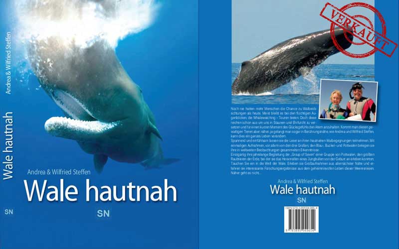 Spannend und einfühlsam lassen sie die Leser an ihren hautnahen Walbegegnungen teilnehmen. Mit einmaligen Aufnahmen, vor allem von den drei Großen: den Blau-, Buckel- und Pottwalen, belegen sie ihre in weltweiten Beobachtungen gesammelten Erkenntnisse. Einzigartig ihre jahrelange Begleitung der „Group of Seven“, einer Gruppe von Pottwalen, den größten Raubtieren der Erde, bei der sie das Heranreifen eines Jungbullen von der Geburt an erleben konnten.
 Tauchen Sie ein in die Welt der Wale. Erleben sie Großaufnahmen aus allernächster Nähe und erfahren sie interessante Forschungsergebnisse aus dem geheimnisvollen Leben dieser Meeresriesen.
Näher geht es nicht …
Sachbildband gebunden, 168 Seiten, 231 großformatige Farbabbildungen, Format 28,5 x 23,5,
ISBN 978-3-89594-965-4, Preis 24,90 € - leider ausverkauft und vergriffen!
- info (at) pottwale.de
- +49 173-976 78 23